Predicting Pneumonia from Chest X-rays using Deep Learning, Computer Vision, and Flask
Introduction:
Pneumonia is a respiratory infection that inflames the air sacs in one or both lungs. It can be caused by various pathogens, including bacteria, viruses, and fungi, and is characterized by symptoms such as cough, fever, and difficulty breathing. Timely and accurate diagnosis is crucial for effective treatment, and advancements in deep learning and computer vision are now playing a pivotal role in automating this process.
Understanding Pneumonia:
Pneumonia affects millions worldwide, with a significant impact on public health. Identifying pneumonia in its early stages through medical imaging, such as chest X-rays, is critical for prompt intervention and improved patient outcomes.
Deep Learning and Computer Vision:
Deep learning, a subset of machine learning, involves training neural networks to learn patterns and features from data. In medical imaging, computer vision techniques combined with deep learning models enable the automated analysis of images, allowing for efficient disease detection.
Transfer Learning with VGG16:
Transfer learning leverages pre-trained models on large datasets for a new, related task. VGG16, a widely used convolutional neural network architecture, has proven effective in image classification tasks. By utilizing the pre-trained weights of VGG16, we can harness its knowledge of general image features for our pneumonia prediction model.
from tensorflow.keras.applications import VGG16
from tensorflow.keras import models, layers

# Load VGG16 base model
base_model = VGG16(weights='imagenet', include_top=False, input_shape=(h, w, 3))
Hyperparameter Tuning:
Fine-tuning the hyperparameters is essential for optimizing the performance of our model. Parameters such as learning rate, dropout rate, and the choice of activation functions can significantly impact the model's ability to generalize well.
model = models.Sequential()
model.add(base_model)
model.add(layers.GlobalAveragePooling2D())
model.add(layers.Dense(256, activation='relu'))
model.add(layers.Dropout(0.5))
model.add(layers.Dense(1, activation='sigmoid'))

# Compile the model with Adam optimizer and binary crossentropy loss
model.compile(optimizer='adam', loss='binary_crossentropy', metrics=['accuracy'])
Training and Saving the Model:
Training the model involves exposing it to labelled data (chest X-ray images in our case) and adjusting the weights based on the model's predictions. The trained model is then saved for future use.
# Train the model with the prepared data generators
history = model.fit(train_generator, epochs=50, validation_data=validation_generator, callbacks=callbacks)

# Save the trained model
model.save('pneumonia_prediction_model.h5')
Setting Up Flask for Prediction:
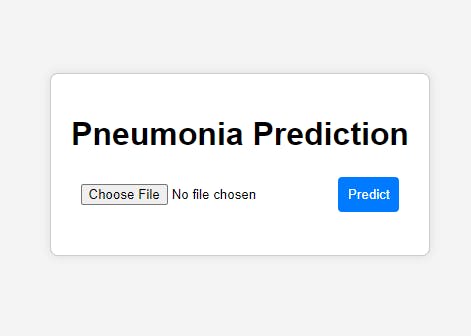
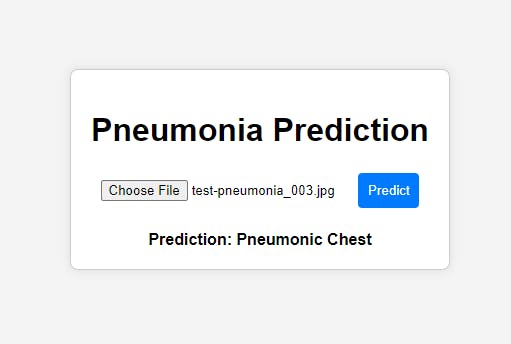
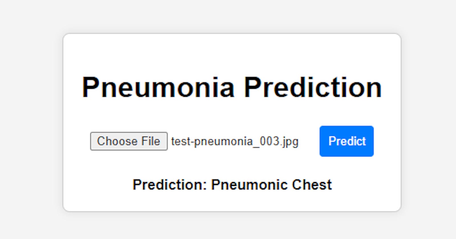
Flask, a web framework for Python, provides an excellent platform to deploy our pneumonia prediction model. By creating an endpoint for predictions, we enable users to upload chest X-ray images and receive predictions in real-time.
from flask import Flask, render_template, request, jsonify
import tensorflow as tf
import numpy as np
import cv2

app = Flask(__name__)

model = tf.keras.models.load_model('model.h5')

def preprocess_image(file):
    img = cv2.imdecode(np.frombuffer(file.read(), np.uint8), cv2.IMREAD_COLOR)
    img = cv2.cvtColor(img, cv2.COLOR_BGR2RGB)
    img = cv2.resize(img, (224, 224))
    img = img / 255
    img = np.expand_dims(img, axis=0)
    return img

def get_class_label(predictions):
    class_index = 1 if predictions>0.5 else 0
    class_labels = ["Normal Chest", "Pneumonic Chest"]
    return class_labels[class_index]

@app.route('/')
def index():
    return render_template('index.html')

@app.route('/predict', methods=['POST'])
def predict():
    if 'file' not in request.files or request.files['file'].filename == '':
        return jsonify({'error': 'No file selected'})

    file = request.files['file']
    img_array = preprocess_image(file)
    predictions = model.predict(img_array)
    class_label = get_class_label(predictions)

    return jsonify({'prediction': class_label})

if __name__ == '__main__':
    app.run(debug=True)


Conclusion:
In this article, we explored the application of deep learning and computer vision in predicting pneumonia from chest X-rays. Leveraging the power of transfer learning with the VGG16 architecture, we built a robust model capable of accurate predictions. The model was then trained, saved, and integrated into a Flask web application, providing a user-friendly interface for real-time pneumonia predictions. This combined approach showcases the potential of cutting-edge technologies in automating medical diagnostics for improved patient care.

